REMS vs. “That Prague Paper”: What the Data Really Says

live better, longer

November 21, 2025
A new paper claims REMS is driven by age, sex, and BMI—not bone. Dr. Doug breaks down the methods, shows why scanning a post-op hip is invalid, and reviews the larger evidence base behind REMS accuracy.

Quick refresher: why REMS matters

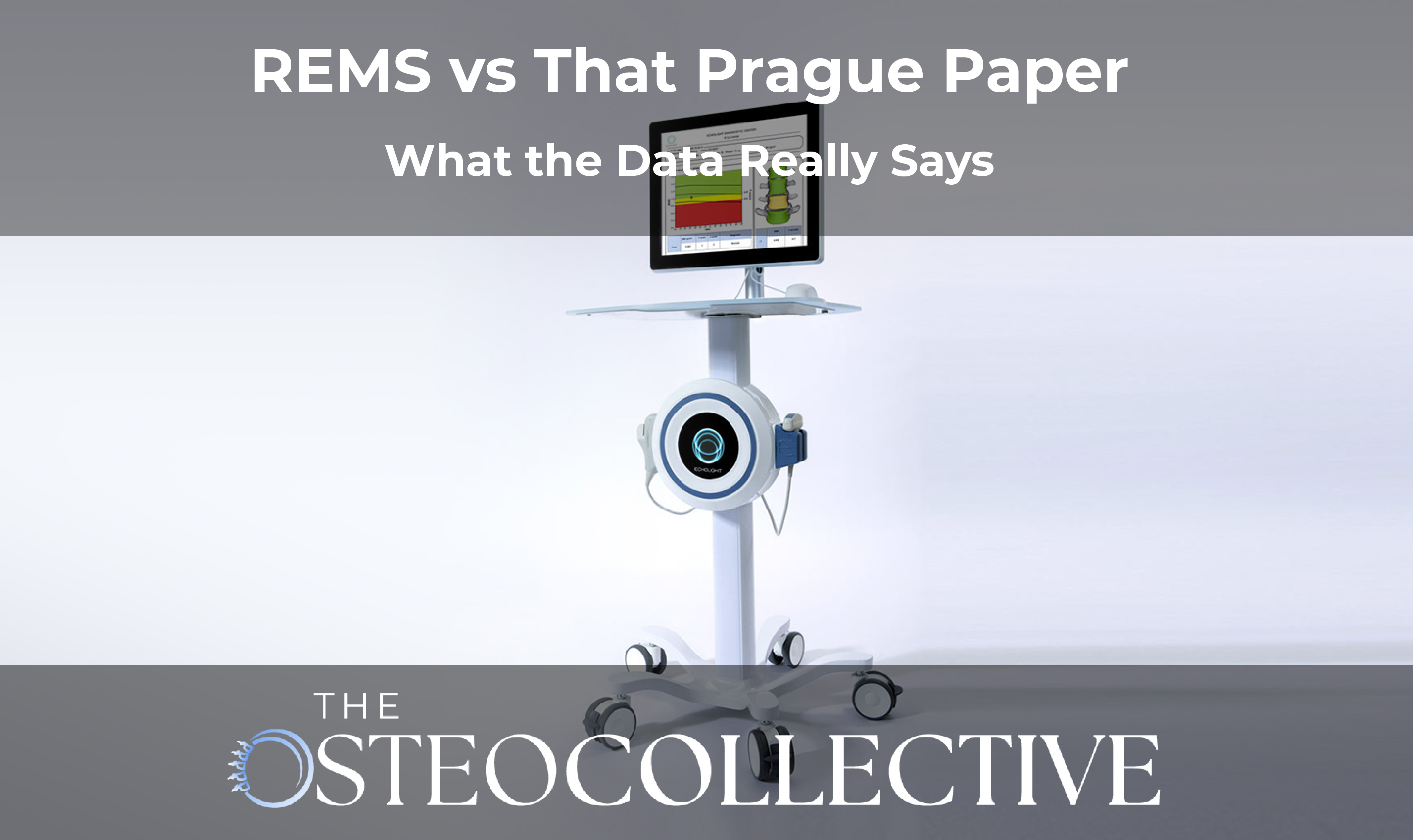
REMS (radiofrequency echographic multi-spectrometry) is a portable ultrasound-based method that reports both density and quality signals from bone. In real life, that means fewer of the positioning and artifact pitfalls that can skew DEXA—especially in people with degenerative changes, vascular calcifications, or spinal hardware.

I’ve recommended REMS for years because its physics (analyzing the raw backscatter spectrum) give it an edge where DEXA often struggles. So when a recent Osteoporosis International study claimed REMS results were mostly driven by age, sex, and BMI—not bone—I dug in.

What the Prague study actually did

  • Design: 50 patients (men + women) with severe hip arthritis scheduled for total hip replacement (THR).

  • Protocol: Measured femoral neck with REMS before surgery, then three days after surgery—when the native femoral neck had been removed and replaced with metal.

  • Finding: Average “BMD” dropped ~2.8% post-op. Authors concluded ~90% of the REMS output could be explained by demographics (age/sex/BMI), not bone.

  • Sub-study: In 7 patients, they typed in fake ages/BMIs (e.g., BMI 13 and 43) and saw REMS values shift—arguing REMS “leans on a calculator” rather than a measurement.

Why that interpretation doesn’t hold up

  • You’re not supposed to scan a replaced hip. The Echolight user manual explicitly says do not scan the operated side; scan the contralateral hip. Metal blocks ultrasound—there’s no meaningful bone signal to analyze.

  • Their published “post-op” images don’t look like metal. The pre- and post-op frames look nearly identical, and neither looks like a true femoral neck view (curvature is missing). This suggests they scanned the wrong region (e.g., diaphysis/lesser trochanter)—outside the intended algorithm.

  • Three days after a THR, tissues are inflamed. Post-surgical edema and hematoma distort ultrasound. Comparing a clean pre-op neck to a traumatized field isn’t a valid test of device accuracy.

  • Garbage in, garbage out. Feeding physiologically impossible inputs (BMI 13) to any reference-guided device (including DEXA) will yield nonsense. That doesn’t prove the device ignores physics; it proves the test conditions were unreal.

  • No DEXA comparison. If you’re claiming a diagnostic tool fails, you’d typically benchmark it against the accepted comparator. They didn’t.

“Saying REMS is ‘just a calculator’ because it didn’t behave in a metal-filled, post-op field is like declaring your stethoscope broken because you strapped it to a lamp post.”

The rebuttal from REMS experts

Within days, orthopedic and REMS specialists (Drs. Nick Burch, Kim Zambito, David Tagnerini, and colleagues with the International Institute for Musculoskeletal Health Education) issued a point-by-point reply: incorrect use case (scanning a replaced hip), questionable image targets, unrealistic data spoofing, and no DEXA control. Add in a small, homogeneous sample (arthritic, elective-surgery population—not a low-BMD cohort), unregistered trial, and sparse methods, and the conclusions overreach the data.

Weighing one outlier against the bigger body of evidence

Across the last decade, 100+ peer-reviewed papers (including multi-center trials) show REMS has:

  • Good agreement with DEXA for classification

  • Lower variance and better reproducibility (lower LSC) in artifact-prone situations

  • Clinical utility recognized in 40+ countries (US, Canada, EU, UK, Australia, Japan)

Do we want more, better, longer studies? Absolutely. But a small, methodologically shaky paper doesn’t overturn a decade of converging evidence—or the physics of REMS itself.

Practical takeaways

If you’re a patient

  • Ask for REMS when DEXA is likely artifact-laden (spine OA, calcifications, hardware) or if you want a radiation-free baseline and more frequent follow-up.

  • Use the same device, same site, same operator posture for follow-ups; consistency improves reliability—REMS and DEXA.

If you’re a clinician

  • Follow the user manual: don’t scan operated hips; use the contralateral side.

  • Pair imaging with bone turnover markers (e.g., P1NP and CTX) to track directionality over months, not just years.

  • Document positioning, landmarks, and device settings—and treat implausible demographics as data entry errors, not “signals.”

Want help building a bone plan that’s more than a scan?

If you still have questions about which imaging to use—and how to tie it to protein targets, impact/simulated-impact training, and bone-savvy HRTjoin our free masterclass and bring your questions to the live Q&A. Or step into the OsteoCollective community for coaching, templates, and accountability so you actually implement the plan.

Remember: a diagnosis of osteoporosis isn’t the end—deciding to reverse it is the beginning.

JOIN OUR NEXT 
FREE MASTERCLASS
Click Her To Join Today
Most Recent Blogs